3D printing is making a profound difference at the University of Malaya’s Centre for Biomedical and Technology Integration (CBMTI). CBMTI uses Stratasys’ PolyJet 3D Printing technology to deliver a range of services including custom medical implants, prototypes for new devices and patient-specific models for surgical planning. But perhaps the greatest impact is in creating sophisticated training simulators for clinical procedures.

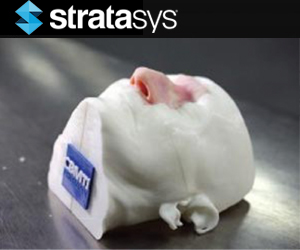
Ear, nose and throat simulation model in three parts.
At CBMTI’s inception, neurosurgeons were mentoring on live human cases, and practicing on cadaveric dissection and computer simulations. Its first 3D printer made spatially accurate models in a single material, but did not mimic human pathology without a costly, time-consuming post process. This changed when they acquired Stratasys PolyJet 3D printing technology.
“Once we got the Stratasys multi-material 3D Printer, we were able to 3D print medical models that could, for instance, mimic the texture of the nose, the linings, and the harder tissue at the back of the nose. We found this very useful, especially in teaching trainees how to handle different materials,” said Vicknes Waran, MD, director of CBMTI.
Better Prepared with Realistic Patient-Specific Models
CBMTI now 3D prints detailed multi-material models that mimic real anatomy, even down to a specific patient’s tumor. With access to advanced multi-material 3D printing, CBMTI can fabricate models that feature different textures and densities over surfaces and throughout interiors, just as human body parts do.
“The J750 allows us to create models with both texture and color variations that mimic actual tissue handling and appearance better for these complex models,” said Dr. Waran. “With the Connex, we can simulate realistic layers of human tissue like skin, bone, dura, brain and tumors within the 3D printed medical model for surgical simulations.”
Customizing the Training Model: How It Works
CBMTI develops its training courses in partnership with leaders in various fields. Together, they identify a patient with the anatomy and pathology they wish to train physicians to treat. CBMTI engineers then convert the patient’s CT and MRI scans into digital design files, and select materials that best match the physical, tactile and color characteristics of the target anatomy. CBMTI has even found ways to use support material, typically removed from the final model, to enhance clinical realism.

The simulation section is installed in a reusable 3D printed head fixture.
“We have also incorporated features such as fluid dynamics so we can simulate endoscopic neurosurgical procedures,” said Yuwaraj Kumar Balakrishnan, CBMTI chief operations officer. “We find surgeons who train on these models are much better prepared in terms of dealing with complex surgeries, simply because they are able to train and retrain on the models until they perfect the procedure.”
Learn more by watching the video below.

Leave A Comment